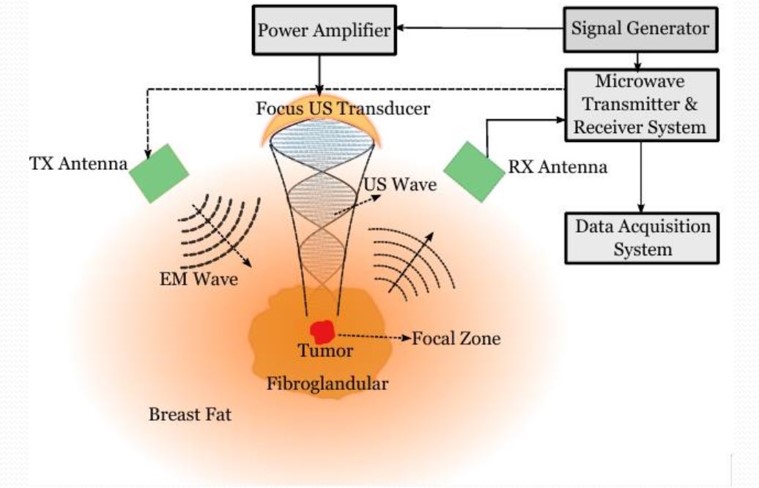
Harmonic Motion Microwave Doppler Imaging (HMMDI)
A hybrid medical imaging method is proposed for imaging using dielectric and elastic properties of the body tissues. Local harmonic motion is generated inside the tissue using a focused ultrasound probe. A narrow-band microwave signal is transmitted to the tissue. The Doppler component of the scattered signal, which depends on the dielectric and elastic properties of the vibrating region, is sensed. The effects of operating frequency, antenna alignment and distance, and tumor depth on the received signal were studied. The effect of harmonic motion frequency on the vibration amplitude and displacement distribution was investigated with numerical studies. The safety of the method was also analyzed in terms of microwave and ultrasound exposure.
Experimental systems were used to obtain HMMDI images from tissue-mimicking phantoms at multiple vibration frequencies. The developed phantoms mimicked the elastic and dielectric properties of breast fat tissue with millimetric-sized tumor inclusion(s). Two-dimensional HMMDI images of the phantoms were obtained by scanning a focused ultrasound probe in two lateral directions. The inclusions inside the fat phantom were resolved showing the potential of the method.